We can distinguish:
-
Mobile and ubiquitous salmonella infections, known as paratyphoid infections, which are a public health problem, and in very rare cases pathogenic in poultry. These are mainly Salmonella Enteritidis (SE) and Salmonella Typhimurium (ST).
-
Immobile and strictly avian salmonella infections by Salmonella Gallinarum-Pullorum (SGP). In animals, we speak of pullorosis or typhoid, depending on the case.
-
Mobile salmonella infection, by Salmonella arizonae, which cause clinical signs, especially in turkeys.
The disease agent and its pathogenicity
The current nomenclature classifies SGPs as: Salmonella enterica, subspecies enterica, serovar Gallinarum (SG) or Pullorum (SP). We now consider that there is only one serovar: Gallinarum-Pullorum, but with two different biotypes (this is still a matter of debate). For simplicity, SP is considered to be a pullorosis agent and SG is considered to be a thyphosis agent.
These are Gram -, facultative anaerobic bacilli, which can be grown at 37°C on Mc Conkey medium (MCK), on blood enriched Columbia agars (COS) but also on more specific media, such as XLD, XLT4 and brillant green agar.
The mechanisms have not been formally identified, but intestinal lesions appear to be the entry point. Salmonella can also invade mononuclear phagocytes, which is not the case in ducks, which very rarely develop salmonellosis.
Epidemiological data
SGPs have a global distribution, but thanks to eradication programmes their prevalence has become almost zero, except in some countries.
Chickens are the natural host of SGPs, but infections are possible in turkey, guinea fowl, quail and pheasant.
The transmission can be horizontal, vertical and pseudo-vertical. It is done via faeces, by contamination of bedding, water and food. Vertical contamination involves contamination of the ova, and pseudo-vertical transmission involves contamination of the shell that causes infection at the time of hatching. An infected hen can contaminate more than 30% of its entire egg production with this process.
After clinical infection, animals can become healthy or asymptomatic carriers. Mortality is highly variable and can reach 100% in some cases. Morbidity is always higher than mortality because some animals can heal spontaneously.
Clinical manifestations of the disease
Two clinical forms are considered depending on the age of the affected animals. SP pullorosis usually affects chickens 2 to 3 weeks old. SG avian typhoid mainly affects adults, but youngs are sometimes affected.
Pullorosis: When eggs are infected, the first clinical signs may appear during or just after hatching. Weakness, drowsiness, decreased appetite, difficult growth and relatively rapid mortality are observed. We can also have dead animals in the shell during incubation. Signs and mortality can also appear during the 2nd and 3rd week of life. Chicks gather under the radiants, have liquid faeces, may have tibiotarsal arthritis, respiratory signs and sometimes blindness. Survivors are delayed in their growth and become carriers later on.
Typhosis: Animals show a decrease in shape, dishevelled plumage, liquid faeces and may in some cases suffer a fall in egg laying, decreased hatching and fertility in breeding animals. There is a decrease in food consumption. Death appears in 5 to 10 days. An increase in body temperature can be detected.
For typhoid and pullorosis, turkeys are anorexic, adypseic and apathetic. They tend to isolate themselves. They have droppings that are liquid and can turn yellow to greenish. Death can occur without clinical signs but it is always accompanied by an increase in body temperature.

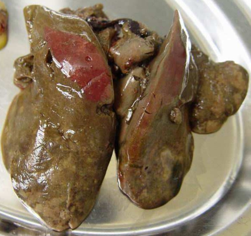
At autopsy, hypertrophy of the spleen and liver is observed, especially in youngs. There are sometimes necrotic foci on the liver, but also on the heart and lungs (in case of respiratory signs). Necrotic foci in turkeys are also found in the mucous membrane of the intestine; they are observable by transparency before opening. Splenomegaly and necrosis areas on the heart are also found in turkeys. The yolk of chicks may have a fibrinocaseous appearance. In case of lameness, a purulent exudate is found in the joints. At the genital level, atrophy of some ovarian follicles is observed. Caseous salpingitis, pericarditis and fibrinous peritonitis may also develop.
The diagnosis
Laboratory diagnosis
- Bacteriology : it is the exam of choice. The liver, spleen and caeca are taken, but also other injured organs such as the heart, lungs, ovary, yolk… You can sow on trypticase soy agar enriched with sheep blood (small translucent colonies) then make a Gram stain and a biochemical identification. A fresh state and a mannitol-mobility test highlight the mobile nature of the isolate.
- Serology : it is useful when the herd shows few clinical signs. The very fast and widely used slide agglutination test has high rates of false positives (interference with vaccination against Salmonella Enteritidis). It may be preferred the serogglutination on a tube, slower.
Differential diagnosis
Lesions are not pathognomonic. We can think of other salmonella (liver, spleen, intestines), aspergillosis (lungs), Mycoplasma synoviae, Staphylococcus aureus, Pasteurella multocida, Streptococcus spp and Erysipelothrix rusopathiae (joints), Marek’s disease, hepatitis with inclusions (liver).
Disease prevention and control
Eradication
Tis is the best method. It has been done successfully in a majority of countries. SGP is sensitive to most disinfectants commonly used in poultry farming and is destroyed by heat at 65°C for several days (manure composting). As virulent materials are in the excreta, care must be taken when spreading droppings and manure, as well as when transporting them. In addition, insect control is necessary because insects are significant vectors. The same applies to rodents. In general, particular attention will be paid to biosecurity conditions.
Vaccination
It is not compatible with eradication measures. However, table egg laying hens are vaccinated in some countries with a SGP vaccine to control Salmonella Enteritidis.
Treatment
If clinical signs appear and the disease has already started to progress, antibiotic treatment (sulfonamides, nitroflurans, tetracyclines, chloramphenicol) can be started. However, these treatments reduce clinical signs but are not sufficient to clean up a batch.


