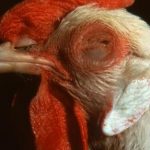
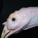
Avian metapneumoviroses are respiratory diseases, described since the 1970s. They include 2 diseases with identical symptoms and lesions : turkey infectious rhinotracheitis and infectious swollen head syndrome.
The disease agent and its pathogenicity
Metapneumoviruses belong to the paramyxovirus family. Subgroups A, B, C and D are distinguished. These are enveloped single-stranded RNA viruses with negative polarity. Their shape and size are variable : spherical to filamentous, from 80 to 1000nm.
Subgroups A, B and D (D in France) are found in turkey infectious rhinotracheitis and infectious swollen head syndrome cases, and subgroup C (Colorado) has been identified in turkey infectious rhinotracheitis cases in the United States. The DuPV variant (Duck Pneumovirus) belongs to subgroup C and is responsible for egg-laying falls and respiratory disorders in palmipeds. There is a greater similarity between human and avian metapneumoviruses in subgroup C than between aMPVs in subgroups A, B and D compared to subgroup C. In addition to turkeys and chickens, there are also metapneumovirus infections in pheasants and guinea fowl.
These metapneumoviruses appear to be resistant to dehydration, and can survive in droppings and manure. However, they are sensitive to common disinfectants (quaternary ammoniums, glutaraldehyde, oxidants).
After incubation for 2-3 days, the virus multiplies in the cells of the ciliated epithelium of the 1st respiratory tract (nasal cavity, trachea). This causes cilia loss and cellular destruction of the mucous membrane of the upper respiratory tract, as well as inflammation. Antibodies are detected 7 days after the start of the clinic, for about 2-3 months. There is a clear relationship between the level of circulating antibodies detected and protection.
Epidemiological data
Sensitive species are mainly hens and turkeys, guinea fowl, and experimentally pheasants, possibly Muscovy ducks. Geese and pigeons seem to be non-sensitive.
The disease can affect any type of production, at any age. But it is more frequent between 3 and 12 weeks in flesh-bearing birds, and at the beginning of egg laying in breeding birds.
Sources are formed by birds emitting virulent materials. The transmission is only horizontal by direct or indirect contact. The virus enters the body by respiratory, ocular and oral routes, by inhalation of viral particles suspended in the air or by contaminated drinking water. Density seems to be a determining factor. Replication of the virus is possible in wild birds, but their actual role in its spread remains to be determined.
Viruses multiply in the epithelial cells of the epithelial and genital tract. In particular, they cause deciliation phenomena in the mucociliary escalator.
Clinical manifestations of the disease
The incubation period lasts 2 to 3 days.
Symptoms of turkey infectious rhinotracheitis
Morbidity is close to 100%, with low mortality (up to 50%).
- In broiler turkey : cough (which can cause oviduct prolapse), sneezing, nasal discharge, tearing and more or less swelling of the periorbital sinuses ; animals show a decrease in behaviour and a decrease in water and food consumption ; the disease lasts about 7-10 days. There is a progression towards healing, or especially after 6 weeks of age, bacterial complications or by mycoplasma (mortality is then higher), or growth retardation.
- In breeding turkeys : in the farm, if the breeding conditions are good, there are often no signs. In laying, there is coughing, discarding, a 15% decrease in egg laying over 10 days appearing 2 days after the respiratory signs. We can have a decrease in the quality of the shell.
Lesions of turkey infectious rhinotracheitis
- In turkey : There are lesions of rhinitis, sinusitis, tracheitis with congestion and abundant mucus. Liver and spleen are enlarged. In the absence of complications, the mucus is clear. During bacterial complications, the mucus is purulent and there are associated lesions of purulent polyserosities : sinusitis, aerosacculitis, splenomegaly, perihepatitis, pericarditis. Ovary and oviduct can be atrophied, eggs malformed ; one can have peritonitis.
Cytoplasmic eosinophilic inclusions are seen in the ciliated cells of the trachea during the first 3 days, followed by non-specific inflammatory lesions.
Symptoms of infectious swollen head syndrome
- In chicken : Morbidity is 4% with mortality below 2%.
Therre are weak moans on chickens 3-6 weeks old, a slight nasal discharge, tearing. Then, we see the appearance of swelling of the head (eyelids, periocular area, sinus, lower jaw, neck), foamy tears and nervous disorders (otitis). There is also a decrease in food consumption. In laying, there is a 5-30% drop in egg laying, evolving over 2-3 weeks, and a decrease in the quality of the shell. - In guinea fowl : The 2 forms : turkey infectious rhinotracheitis like turkey, or infectious swollen head syndrome like chicken.

Lesions of infectious swollen head syndrome
- In hens : There is peri-ocular edema, sinusitis with or without edema, and moderate lesions of rhinitis, laryngitis and tracheitis. Complications include purulent conjunctivitis, ear infections, maxillary arthritis, osteitis, aerosacculitis, pericarditis and perihepatitis.
The histological lesions are similar.
Le diagnostic
Serological
2 blood tests at 15 day intervals, the first as soon as possible.
Virological and PCR
As soon as possible isolation from sinus, nostrils or ocular conjunctival swabs. It is difficult to detect the virus in animals with severe signs. RT-PCR available on a routine basis. It differentiates between serotypes A, B and C. Viral isolation is mainly used in research.
Differential diagnosis
Infectious bronchitis, Newcastle, influenza, mycoplasma infections.
Disease prevention and control
The treatment consists of a fight against the germs of complication.
Prevention is first and foremost a matter of health, with the importance of a single strip and reasonable density, compliance with biosecurity rules and good animal husbandry practices.
Due to the high contagiousness of the disease, medical prophylaxis is recommended. Live virus vaccines are available for vaccination of young birds, and inactivated virus vaccines are available for use on breeding stock before laying.
Vaccination has 3 objectives : to obtain the earliest possible active protection, to have the longest possible protection, and to generate significant local immunity in the respiratory mucosa.
To do this, vaccination takes place in the first week of life, or even at hatching, 1 or more booster doses are carried out, and vaccination is carried out by spraying.
