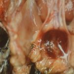
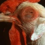
Marek’s disease is a lymphoma of viral origin, associated with nerve or visceral tumors. The first description dates back to 1907 in Hungary, by Professor Marek. This disease really emerged as a major constraint to global poultry production in the 1960s, with the emergence of pathogenic variants. Since then, the spread of vaccination has made it possible to control this infection relatively well : accidents, often linked to poor vaccination practices or particularly pathogenic isolates are regularly observed.
The disease agent and its pathogenicity
The agent of Marek’s disease is a herpesvirus (Marek Disease Virus : MDV). It is a “large” enveloped virus, whose genome is a large double-stranded DNA. Classically, 3 serotypes are distinguished :
- Serotype 1, the only pathogen in chicken. More or less virulent strains are described, with the emergence of “very virulent” viruses : vvMDV, even vv+MDV…
- Serotype 2, non-oncogenic (the SB1 vaccine strain is used in the USA)
- Serotype 3, or turkey herpesvirus (HVT : Herpesvirus of Turkey): non-oncogenic, used as a heterologous vaccine against serotype 1.
The taxonomy of these viruses has been updated :
- Marek’s disease virus (serotype 1) is called gallidherpesvirus 2.
- Serotype 2 is called gallidherpesvirus 3.
- The turkey herpesvirus is called meleagridherpesvirus 1.
The Marek virus can take 2 forms :
- Form integrated into the host cell genome : associated with the tumor process, this form is non-infectious.
- Free form at the subepidermal layer in the process of desquamation of the feathery follicle. It is the contaminating form, released into the external environment.
The pathogenic process of Marek’s Disease can therefore be summarized as follows :
- The contamination of a naive chicken is mainly by breathing infectious scales, with replication in pulmonary macrophages.
- Diffusion into lymphoid tissues (Fabricius bursa, thymus, spleen), then viremia which can lead to 2 types of infection :
- Lytic infection in the Malpighi epithelium of feathery follicles (contaminating scales) : this process explains the general spread of the virus in the environment (epidemiological importance)
- “Restrictive productive” infection (infection is strictly associated with cells) of T lymphocytes : transformation and development of tumors : it is the latter process that causes clinical manifestations (clinical importance).
The development of these tumors is slow, which explains why Marek’s disease mainly affects animals with a long economic life (“label” chicken (high quality chicken), laying and breeding hens). Chicken is the susceptible species, turkey can be affected (especially Christmas farm turkeys with a particularly long economic life). It should be noted that infection is universal, even after decades of vaccination. Some chicken strains have a higher genetic resistance ; it appears that MHC1 haplotypes play a role in this resistance (especially B21B21 haplotype).
The virus is transmitted only horizontally (scales), directly and indirectly, mainly by the respiratory route. The virus protected in desquamated cells is highly resistant in the external environment. (Several months at a temperature of 20-25°C, several years at 4°C).
Clinical manifestations of the disease
Clinical signs of MM usually appear at about 3 weeks of age and the peak of infection occurs between 2 and 7 months of age.
Symptoms
Birds may have asymmetric lymphoid infiltration of the peripheral nerves leading to partial paralysis and/or dilation of the crop due to paralysis of the vagus nerve. GaHV-2 can also infect the brain, resulting in transient paralysis or persistent neurological disease. Blindness is associated with lymphoid infiltration of the iris. Birds with visceral tumors can appear depressed and are often cachectic before death.
The cutaneous form (which was called “cutaneous leucosis”) is located at the feathery follicles. Nodular lesions may involve a few dispersed follicles or they may become coalescent, and redness of the skin is often noted.
Lesions
Lymphomas can affect a wide variety of tissues :
- Nerve damage : brachial plexus, lumbosacral, sciatic nerve
- Tumor damage to the liver, ovary, testis, spleen, heart, muscles
- Skin lesions : enlarged hair follicles
Histologically, there is diffuse or focal mononuclear cell infiltration.


The diagnosis
The main methods for diagnosing the disease are based on :
- the detection of lymphomatous infiltrates by histopathology
- detection of DNA (PCR) or viral antigens (immunohistochemistry)
- real-time quantitative PCR allows to discriminate wild strains from Rispens or HVT vaccine strains and to detect the virus in feathery follicles in particular
NB : the detection of antibodies is not operative because the presence of antibodies is in no way related to the disease, infection by non-pathogenic viruses being very frequent. Isolation of the virus by cell culture remains a field of research.
The diagnosis is first based on anatomo-pathological analysis, in particular microscopic analysis :
- If nervous infiltration + / viscera + : MAREK
- If nervous infiltration + / viscera – : MAREK
- If nervous infiltration – / viscera + : (MAREK)
- If nervous infiltration – / viscera – : NOT MAREK
Differential diagnosis
Must be made with avian leucosis (retroviruses), which are generally much rarer. The keys to this diagnosis are summarized below :
| Marek’s disease (herpesvirus) | Leukemia (retrovirus) | |
|---|---|---|
| Age of onset of the disease | After 6 weeks | Rare before 14 weeks Often around 30 weeks |
| Morbidity | + than 5% | – than 5% |
| Symptoms | Frequent paralysis | Non-specific |
| Nerves hypertrophy | +++ | No |
| Gonadal tumors | +++ | Rare |
| Eye tumors | + | No |
| Skin tumors | + | No |
| Liver, spleen, kidney tumors | + | +++ |
| Fabricius bursa tumors | Rare | +++ |
Disease prevention and control
Compliance with biosecurity principles must be recalled, but the universal presence of the virus makes vaccination essential in animals with long economic lives. In any case, hygiene makes it possible to limit early contamination before post-vaccination protection.
Vaccination is carried out on the 1-day-old chick, or even in ovo.
There is no point in vaccinating older animals !
In France, 3 types of vaccines are used :
- Lyophilized HVT serotype 3
- Frozen HVT serotype 3
- Frozen attenuated strain serotype 1 (Rispens) The Rispens strain (or CVI-988) is widely used in France.
Warning: Marek vaccination does not protect against infection but only against the development of tumors.
It does not prevent infection, and therefore the spread of the wild virus in vaccinated animals, even after years of systematic vaccination on a farm.
Vaccination practices must be followed :
In case of use of frozen vaccine : the thawing time -resumed in the diluent- administration of the vaccine is crucial : the viral titer decreases very quickly, hence the need to administer the vaccine as soon as the dilution is made.
Quality of the injection : check the adjustment and maintenance of the vaccination machines, and especially the respect of good practices by the staff of a repetitive act (2000 to 3000 chicks/hour).
NB: in ovo vaccination, carried out at 18 days (transfer from the incubator to the hatchery) using an automatic system, is spread in the hatcheries : it makes it possible to vaccinate from 30 to 50,000 eggs/hour.
