Histomoniasis is a parasitic, infectious disease specific to galliformes. It is a typhoid hepatitis that mainly affects turkey and guinea fowl. This disease has been described for a long time (since 1895). It had become very rare since the use of effective pest control products until the early 2000s. The ban on anti-histomonic drugs (2003) led to a re-emergence of the disease, mainly in the turkey chain.
The disease agent and its pathogenicity
The causative agent is Histomonas meleagridis, a 10µm polymorphic flagellate protozoan.
Two forms exist in the definitive host : a form without flagellum in the tissues, and a form with flagellum in the caecal light.
The evolutionary cycle of Histomonas meleagridis is, in part, linked to the cycle of the nematode Heterakis gallinarum, a parasite of poultry caeca. Transmission of H. meleagridis from one host to another occurs via resistant H. gallinarum eggs in the external environment. The eggs ingested release the protozoan into the caecal light, where it multiplies. Then it passes through the caecal wall into the blood and into the liver. In the caeca, it cohabits with adults H. gallinarum, which it can reinfect. H. gallinarum eggs ensure the survival of H. meleagridis in the external environment, but also protection in the digestive tract. Parasitized eggs of H. gallinarum can be ingested by earthworms that accumulate and carry larvae carrying H. meleagridis.
While it has long been considered that H. gallinarum plays an essential role in the parasitic cycle of H. meleagridis, another possibility of direct transmission of the trophozoic form by ingestion of infested faeces (“cloacal dropping“) is now accepted.
Epidemiological data
Many galliformes host the parasite. Turkey, guinea fowl and partridge are very sensitive, while hens, pheasants or quail express a more discreet form (serious clinical episodes are reported in laying hens, however).
Currently, the species most affected by clinical forms of histomoniasis is turkey. There are also differences in sensitivity between poultry strains.
The most serious forms in turkeys are expressed at the end of the first month of life, but especially between 8 and 18 weeks of age. In hens, symptoms are visible at 3 to 5 weeks of age. Older hens and turkeys are susceptible but clinical forms are rarer or less marked. However, they do excrete many Heterakis eggs.
Low-sensitivity poultry (hens), old turkeys, and wild birds are healthy carriers that constitute a reservoir.
Heterakis plays a significant role as a vector, as it parasites the same hosts as H. meleagridis, and protects it in the external environment. The earthworm has a role in the spread and conservation of parasites.
Clinical manifestations of the disease
The incubation period is 7 to 10 days.
Symptoms
One of the first signs of histomoniasis is sulphur yellow or mustard diarrhea, a sign of a caseous inflammation of the caeca. Other signs are feathers stained with droppings, anorexia, prostration, abnormal gait and head down or hidden under a wing. A dark coloration of the head (“blackhead disease“) can also be observed. Birds become very thin.
Mortality can be high (up to 80%) and persistent. It can be amplified by secondary infections. Survivors will show growth retardation.

Lesions
Lesions are early and precede symptoms. They mainly concern caeca and liver.
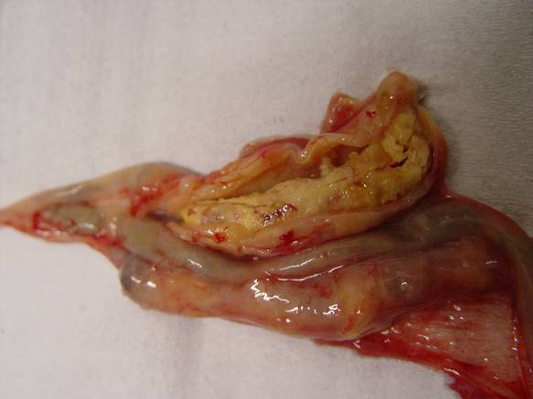
- Caecal lesions involve one or two caeca, all or part of the caecum. The caecal walls are thickened and congested, with abundant exudate distending the caecum. The caeca then look like irregular, firm sausages and have a thickened wall. At the opening, ulcerative and necrotic lesions are observed, with a caseous plug. A possible evolution is the perforation of the caecum which causes abdominal serositis. In chronic forms, adhesions between the caecum and the intestine, or concerning the abdominal serosa, are observed.
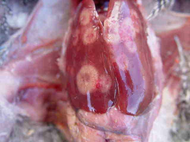
- Liver damage is less frequent and more variable. An enlarged and discoloured liver can be observed. But the classical lesions are circular necrotic foci, with the appearance of a targetoid spot in depression, with raised edges. These stains measure from a few millimeters to a few centimeters, and give the liver a characteristic appearance.
Other organs (kidneys, lung, spleen) may also have round necrotic foci.


The diagnosis
Epidemio-clinical diagnosis
- Lésions d’autopsie : lésions caecales avec boudin caséeux, foie tacheté avec cocardes en dépression
- Epidemiology : epizootic on young animals, turkey
- Symptoms : yellow diarrhea, anorexia, prostration, gait, black head
- Autopsy lesions : caecal lesions with caseous “sausage”, spotted liver with depressed cockades
Laboratory diagnosis
The diagnosis of certainty is based on the detection of the parasite by direct examination under the microscope. This technique is not easily achievable and requires a quick examination after sampling.
Cultivation is possible but requires the use of a specialized laboratory.
A PCR technique has been developed at the ENV in Lyon but is not routinely available.
Differential diagnosis
Avian tuberculosis, Salmonellosis, Pasteurellosis, Marek’s disease, caecal Trichomoniasis, caecal Coccidiosis with E. tenella, …
Disease prevention and control
Several molecules are effective against H. meleagridis and were used for curative or preventive purposes :
- Nitroimidazoles : dimétridazole, ipronidazole, ronidazole…
- Nitrofuranes : nifursol
However, all these specialities have had their marketing authorisation withdrawn as part of the European re-evaluation of maximum residue limit (MRL) dossiers : they are therefore no longer available in the European Union. However, molecules remain available in other parts of the world, for example in the United States or in some Maghreb countries (Histostat®).
In the absence of any effective medical solution, health prophylaxis has become essential. It is important to separate species, especially chickens and turkeys. Heterakis must also be controlled by regular worming of birds.
Different alternative medical approaches (essential oils, homeopathy, …) are tested in the field, without their effectiveness being clearly established.
In the event of a severe outbreak, total slaughter of the batch is sometimes the only economically realistic solution…


