Derzsy’s disease, first described in 1967 by the Hungarian Domokos Derzsy, is a pathological dominant disease in domestic palmipeds. Its distribution is global and its economic impact is major in countries where palmipeds are raised. Several clinical forms of this disease can be observed, depending on the host species, its age, its immunity.
The disease agent and its pathogenicity
The causal agent is a parvovirus, Derzsy parvovirus or Goose parvovirus (GPV). No antigenic relation has been demonstrated with other animal parvoviruses. Similarly, GPV is antigenically and genetically distinct from the Muscovy Duck parvovirus (see specific sheet). Recent genetic analyses have identified three groups of viruses : Hungarian, Asian and West European.
The GPV is a small, non-enveloped, icosahedral symmetric, 20-22 nm in diameter virion. Its genome consists of a single-stranded DNA. The GPV is highly resistant to many disinfectants, heat (65°C for 30 minutes) and acid pH (ph3 for 1 hour at 37°C). However, treatment with formaldehyde at 0.5% can destroy the virus.
The GPV replicates in young cells under active multiplication, its replication being carried out in the nucleus. The virus first replicates in intestinal cells and then spreads through the bloodstream and will be able to exercise its pathogenicity in a wide variety of tissues. Natural infection results in the synthesis of neutralizing antibodies, which can be detected long after infection. For the reproductive female duckling, these post-infectious (or post-vaccinal) antibodies are transmitted to the offspring, in which they persist until about 2 weeks of age.
Epidemiological data
- Derzsy’s disease is common to several species of anatidae, including many wild species. In domestic palmipeds, the goose (Anser anser), the Muscovy duck (Cairina moschata) and the Mulard duck (Pekin female Anas platyrhynchos X Muscovy male crossing) are affected. However, the Pekin duck does not develop the disease.
- The most sensitive population is the youngs, with increased and decreasing sensitivity during the first month of life. This may be explained by the increased number of dividing cells in young subjects (link with damage to parvovirus dividing cells). Adult subjects are affected in tissues whose cell populations are highly divided and multiplied. Extrinsic factors can modify the period of sensitivity : animal density, hygiene, environment, various stresses.
- Sources of contamination are infected animals that excrete the virus in the faeces, and many contaminated secondary sources (bedding, sheds, paths, equipment, etc.). Viral shedding may persist for more than one year. Some animals may develop an inapparent form of the disease and form a reservoir.
- The virus is transmitted mainly horizontally, directly or indirectly. Vertical transmission is also present.
- Birds that have survived infection or are subclinical become carriers and can transmit the virus vertically or horizontally. No biological vectors have been currently identified.
Clinical manifestations of the disease
The incubation period will depend on the age of the affected animal. During the first week of life, symptoms appear within 3 to 5 days. The incubation period for these three weeks of life is 5 to 10 days. The older the individual’s age, the longer the time it takes for n the first symptoms to appear (2 to 3 weeks for an adult).
Symptoms
In geese and Muscovy ducks, several forms involving GPV are distinguished according to the age of the host.
- In subjects less than two weeks old, the disease takes an acute form of progression, with apathy, musculoskeletal disorders, diarrhea and mortality of up to 50%.
- In subjects aged two to five weeks, these are early subacute forms, with dyspnea, anorexia and weight loss ; the batch appears heterogeneous.
- In older subjects, there are late forms, with growth retardation, feather loss and low mortality.
- In some cases, complications by secondary germs can give the illusion of a chronic respiratory disease.
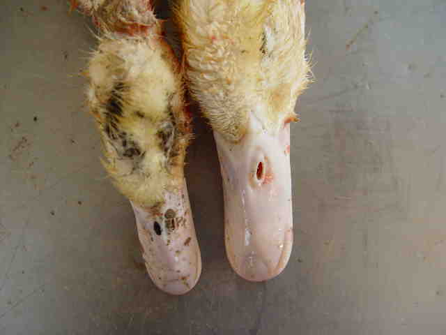
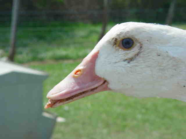
In Mulard ducks, the disease takes the form of short-beaked dwarfism syndrome, with more or less strong manifestations. The clinic is marked by growth retardation, dwarfism, short-beaked animals, “goose profile” animals, spontaneous fractures during handling (especially wings). The affected batch appears heterogeneous, with an attack rate of 10 to 30%.
Lesions
In goose and Muscovy duck
The lesional picture is characterized by a generalized visceral damage, dominated by lesions of ascites, pericarditis, aerosacculitis, nephritis, more or less marked depending on the occurrence of bacterial superinfections. At the histological level, the lesions are found in the heart, liver, splenic, nervous, muscular and tendon tissues.
In Mulard duck
Mostly, lesions of bone tissue are observed : shortened long bones, marked bone fragility, resulting in spontaneous fractures of the humeral and femoral heads.
The diagnosis
Epidémio-clinical diagnosis
In geese and Muscovy, symptoms, age of onset and generalized lesions of visceral damage guide the diagnosis.
In Mulard, the clinic is suggestive of marked short-beak dwarfism syndrome ; in more rough episodes, suspicion is based on the observation of a heterogeneous lot.
Differential diagnosis
In geese : hemorrhagic enteritis nephritis of the goose, riemerellosis, zootechnical errors
In ducks: duck plague, reovirosis of Muscovy, parvovirus of Muscovy, riemerellosis, cholera, zootechnical errors…
Laboratory diagnosis
- Histology : the observation of lesions of degenerative myocarditis and degenerative hepatitis ; and especially, the observation of intranuclear inclusions of the Cowdry A type allow a diagnosis of certainty.
Caution : in subacute forms of evolution, these lesions are very rarely observed, especially in Mulard ducks. - Virology : There are many methods of cultivation. The virus can be isolated from embryonated eggs of Muscovy ducks or geese, from primary cell cultures (fibroblasts) of embryos of geese or Muscovy ducklings, with evidence of a cytopathogenic effect. Other methods, such as immunofluorescence or immunoperoxidase, are reserved for research.
A quantitative PCR test is routinely available in France and allows the detection of viral DNA in many organs within a few days. It also makes it possible to discriminate between wild strains of the usual vaccine strain. - Serology : several tests have been developed, but none are currently routinely available : the reference method for detecting neutralizing antibodies remains serum neutralisation, which is very cumbersome to implement ; an ELISA test recently developed in Hungary should be available in the field soon.
Disease prevention and control
Prevention
In unscathed farm, it consists in preventing the introduction of GPV. Given the resistance of the virus, this requires rigorous measures : monitoring the introduction of animals, separating age classes, …
In contaminated farm, the elimination of the virus involves the removal of animals from the farm, followed by several cycles of cleaning and disinfection of sheds, paths and equipment, using an approved virucide ; a review of farming practices may be recommended, in particular to promote single-band farming.
Vaccination
Several vaccines are available on the French market : live attenuated virus or inactivated adjuvanted virus vaccines. Similarly, a distinction is made between monovalent vaccines (Derzsy) and bivalent vaccines (Derzsy + Muscovy parvovirus).
Four specialities are available in France : PalmivaxÒ (Merial), ParvokanÒ (Merial), DeparmuneÒ (CEVA SA) and ParvolÒ (derogation status).
The vaccination scheme against Derzsy’s disease remains highly debated : it traditionally includes a primary vaccination of future breeders, followed by booster shots before (or even during) egg laying, and the vaccination of ducklings growing at the age of 1 day and/or at 14-21 days. The latter choice depends largely on the vaccination protocol applied to breeding animals and the infection pressure at the start. All these vaccinations are given by parenteral, subcutaneous or intramuscular route.
Maternal antibody transmission is carried out by affected or vaccinated individuals to their offspring. These persist for the first 2 to 4 weeks of life. Vaccination with an inactivated vaccine, rich in antigen, at 1 to 2 weeks of age therefore provides good coverage.
A recombinant vaccine is under development.


