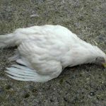
Colibacillosis is probably the most frequent and important bacterial infection in avian pathology. They can lead to mortality, reduced performance and seizures at the slaughterhouse. Unlike mammal infections, avian colibacillosis takes general forms, with a respiratory or genital entryway. Most colibacillosis are superinfections, following viral or bacterial infections (respiratory mycoplasma in particular).
The disease agent and its pathogenicity
The etiological agent of colibacillosis is the bacterium Escherichia coli (E. coli). It is a Gram-, non-sporulating bacterium, of the Enterobacteriaceae family. This bacterium is most often mobile. It is characterized by the antigens O (somatic), H (flagellar), F (pilus) and K (capsular), which allow several serotypes to be identified. In birds, the serotypes “considered pathogenic” are O1K1, O2K1 et O78K80. New pathogenic (non-typable) serotypes are emerging.
The bacterium is sensitive to common disinfectants.
The pathogenicity of E. coli is based on their ability to colonize the respiratory tract, their resistance to the immune system, their ability to multiply in an iron deficiency context, and their ability to produce cytotoxic effects. Several potential virulence factors are identified in avian E. coli : fimbriae adhesins, protein with haemagglutinating activity, aerobactin system for iron capture, polysaccharide capsular antigen, resistance to the bactericidal power of the serum, toxins and cytotoxins.
Impacts on public health
Some pathotypes of E. coli that can infect humans could be carried by poultry. Preventive antibiotic therapy for poultry favors – at least in principle – the pressure to select multi-resistant strains. Antibiotic use should therefore always be reasoned to use antibiotics to which the bacterial isolate targeted is sensitive and according to an appropriate dosing regimen (route, dose and duration of treatment). The use of fluoroquinolones should be reserved for second-line treatments in the event of therapeutic failure.
Epidemiological data
-
All avian species are susceptible to E. coli. It is an extremely common and globally distributed infection. Some factors predispose poultry to the disease.
-
Young age, stress, high ammonia levels, a decrease in temperatures, concomitant infections, all contribute to colibacillosis. In most cases, E. coli should be considered more as a superinfection agent than as the primary cause of a disease.
-
There are several forms of the disease : localized forms, an acute septicaemic form, chronic forms.
-
Young birds are more sensitive to the septicaemic form. Cellulite is promoted by skin erosions and poor bedding. Omphalite is induced by faecal contamination of eggs, broken infected eggs, salpingitis or concomitant ovaritis in the mother. Genital forms are found in future reproductive female before laying eggs or in adults with or without respiratory signs. The venereal form in turkeys occurs after the first artificial inseminations. Respiratory forms are mainly found in young birds, mainly in superinfection.
-
E.coli is a normal bacterium of the digestive tract of poultry. It is therefore spread by the faeces of sick or carrier birds and birds are constantly exposed (by sick or carrier birds, rodents, insects (tenebrous and fly), wild birds, water, dust, environment). As soon as a bird’s resistance is weakened, pathogenic or non-pathogenic strains can develop.
-
E.coli, present in the intestines, nasal tracts, air sacs or genital tract can be a latent source of infection. Some pathogenic strains can also infect an unweakened bird.
-
Contamination is mainly by air tracts through aerosols. Bacteria are inhaled and contaminate air bags. These can prolong the infection to genitals by contact. Some intestinal E. coli cause systemic infections after enteritis. True vertical transmission is possible but rare. Eggs can be contaminated on the surface when they pass through the cloaca or soiled litter.
Clinical manifestations of the disease
Localized forms
- Omphalitis and yolk sac infection : Variable mortality is noted. The umbilicus is edematous and inflamed, with the presence of scabs. The yolk sac is poorly resorbed, with an opacified and congested wall, a greenish to yellowish content. Aerosacculitis and pericarditis are sometimes associated with it.
- Cellulite : Oedema and subcutaneous caseous exudate are observed in the ventral abdominal region, particularly under the thighs. It often follows the presence of skin lesions.
The bird does not show clinical signs, it is often a discovery at the slaughterhouse leading to the seizure of the carcass. It can therefore cause major economic losses.
E. coli is the main etiological agent isolated in cellulite lesions.
There is a form of cellulite located in the head, which begins in the periorbital region (entry of E. coli due to conjunctivitis or other weakening of the ocular mucosa) - Genital forms : Salpingitis and ovaritis : There is a caseous exudate, sometimes lamellar, in the oviduct, often associated with intra-abdominal egg laying. We can have a drop in egg laying and associated mortality.
- Venereal colibacillosis of turkey : This form is often fatal. Caseo-necrotic vaginitis, peritonitis, abdominal egg laying and cloacal and intestinal prolapse are observed.
- Enteritis : The intestines, especially the caeca, are pale and dilated by a liquid content.
Respiratory form
- Birds are indolent and anorexic. They have non-specific respiratory symptoms : wheezing, coughing, sneezing, nasal discharge, sinusitis.
- At the lesional level, inflammatory lesions of the visceral serosa are observed : pericarditis, perihepatitis, airsacculitis, more or less exudative.
Acute systemic form or colisepticemia
- Variable morbidity and (sudden) mortality are observed. The lesions are non-exudative. The liver is enlarged, with some areas of degeneration. The spleen is enlarged with necrosis points. Multiple inflammatory lesions are observed: pericarditis, perihepatitis, airsacculitis, pneumonia, yolk sac infection, arthritis, osteomyelitis, tenosynovitis, etc…
- These forms can have a respiratory, enteric, or other origin.
Chronic forms
- Different forms of lesions can exist : meningitis, endophthalmitis, arthritis, osteomyelitis, tenosynovitis, Meckel’s diverticulum abscess.
- Hjärre disease (or coligranulomatosis) is a particular form, observed in particular in laying hens : whitish masses or nodules are observed in several organs (along the intestines, in the mesentery, in the liver), except in the spleen. Caseous cylinders are also observed in the caeca (not to be confused with histomoniasis or caecal coccidiosis). Mortality can be high.
The diagnosis

Laboratory diagnosis
Bacterial culture is easy to implement. Fecal contamination should be avoided when sampling. Typing the isolate is necessary, but it is not always possible to conclude on the pathogenicity of the identified strain.

Differential diagnosis
Riemerellosis, pasteurellosis, salmonellosis, infectious coryza, avian smallpox, mycoplasmosis ; tuberculosis in the case of Hjärre disease.
Disease prevention and control
Treatment
The treatment is based on antibiotic therapy. The antibiotic susceptibility test is necessary because of the many antibiotic resistance tests observed on field isolates.
If the choice is possible, it is preferable to use molecules such as oral quinolones (nalixidic acid, oxolinic acid, flumequine), oral lincosamides, parenteral aminosides, oral beta-lactamines, tetracyclines.
Prevention
Health prevention is based on the control of risk factors : food and environmental conditions, water quality, and more generally compliance with biosafety rules.
Probiotic (defined) or normal (undefined) digestive flores may also be administered to 1-day-old chicks from adult subjects, on the same principle as the prevention of salmonella contamination.
Medical prevention can also use inactivated or attenuated vaccines administered to breeding animals to protect young chicks with maternal antibodies. Autovaccines are also indicated in specific cases.


