Pasteurellosis is an infectious disease caused by Pasteurella multocida, affecting many bird species. It owes its name to Louis Pasteur, who specified the characteristics of the germ in question, which had been discovered as early as 1879 by Toussaint. The disease is found worldwide, sporadically or enzootically, acutely or chronically as a systemic disease with high mortality and morbidity.
The disease agent and its pathogenicity
Pasteurella multocida is a Gram negative, immobile, encapsulated, extracellular bacterium, both aerobic and anaerobic. The antigenic structure of the bacterium is complex. It is composed of a capsular antigen = K antigen, which masks the wall antigen or somatic antigen = O antigen. The bacterium is very sensitive to UV rays, desiccation, common disinfectants, and can only resist a few days in an outdoor environment.
The classification is complex. First of all, the strains of pasteurella can be separated into three subspecies : P. multocida multocida, P. multociada septica and P. multocida gallicida. All three can be found during avian pasteurellosis but the most frequent remains the first. Other more precise classifications exist. Carter’s classification distinguishes 4 types of K antigens: A, B, C and D (A being the most frequent in poultry farming). The O antigen makes it possible to classify pasteurella under different serotypes, which vary according to the classifications. According to Namioka‘s classification, the O antigen has 12 serotypes (1 to 12) and thus the pasteurella is classified according to the combination of capsular and somatic serotypes. The Heddleston classification, widely used outside France, distinguishes 16 serotypes (1 to 16), but shows no agreement with the Namioka classification (different serotypes according to Namioka may correspond to the same serotype according to Heddleston).
The pathogenesis is complex. It is a toxi-infection, causing an increase in the permeability of capillaries with water disorders, and disorders of energy exchange between cells. The virulence of pasteurella is related to the bacterial strain, but also to other factors (susceptible avian species, inoculation route, environment) and requires the presence of the bacterial capsule. Acute forms are caused by very virulent strains that produce a large amount of endotoxins. The immunity involved is more of a humoral type.
Epidemiological data
Many bird species are susceptible to P. multocida. The disease is mainly found in turkey, duck, goose and chicken. Humans can be accidentally contaminated by a skin lesion. It is a disease of adult or young adult birds, but the disease can appear as early as 4 weeks. Reproducers are more frequently affected. There are many healthy carriers among wild birds.
Many factors favour the onset and development of infection in a farm. Environmental factors are predominant, especially the cold. Pasteurellosis is most common in late summer, fall and winter. The bacterium persists for a long time and easily in cool, moist soils. Stresses, such as declawing, debeaking, vaccinations and force-feeding, are also very favoring.
Transmission is horizontal, indirect (insect, material,…) but above all direct. There does not seem to be any vertical transmission.
Reservoirs of P. multocida can be infected farm birds (chronic carriers or survivors), or wild birds entering the farm, rats, pigs and domestic mammals are also suspected. Virulent materials are oral, nasal and conjunctival secretions. All faeces and dirt from sick birds are contaminating. The bacteria multiplies easily in dead bodies. The penetration route is mainly airborne, but oral, conjunctival and dermal routes are possible.
Clinical manifestations of the disease
Considering the variability of the pathogenicity of the strains and the resistance of birds, pasteurellosis presents a high clinical and lesional polymorphism.
Symptoms
- The highly acute form can be devastating. During less abrupt evolution, there is intense prostration, hyperthermia ; the crest and barbels are purplish. Death occurs in 3 to 6 hours.
- The acute form is accompanied by hyperthermia, anorexia, dishevelled feathers, tremors, rapid and noisy breathing, discharge ; the crest, barbels and feathered areas are cyanosed just before death. We also have abundant, smelly, greenish diarrhea that becomes hemorrhagic. Some birds may have a stiff neck or vomitings. Death occurs in 2-8 days. If animals survive these initial signs, they may nevertheless die from the debilitating effects of dehydration, present a chronic form or recover.
- In the chronic form, the signs vary according to the location of the infection : pasteurellic abscesses (arthritis, wattle’s disease in chicken), pharyngitis, conjunctivitis, middle ear infection (with torticollis in turkeys), respiratory form (most frequent manifestation resembling chronic respiratory disease with tracheal rams and dyspnea). This form always appears after an acute phase.
Lesions
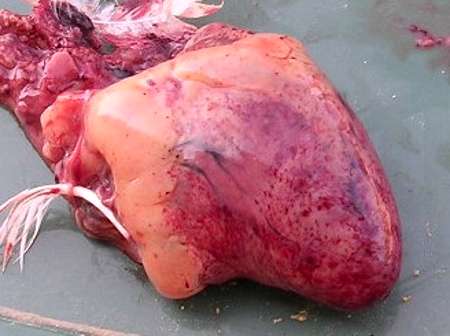
- In the highly acute form, there are non-specific lesions of hemorrhagic septicaemia : generalized congestion, hemorrhagic lesions (especially on the gizzard, heart, small intestine, lungs, kidneys and spleen). Hemorrhagic exudate is also observed in the pericardial and peritoneal cavities.
- In the acute form, some lesions are added to the septicemic lesions : congested liver with a hemorrhagic and then yellowish white staining, pneumonia lesions with yellowish necrosis foci in the pulmonary parenchyma, especially in turkeys and ducks. Other organs may be affected, such as the intestine (fibrinous enteritis), crop and pharynx (mucosal deposition) or ovarian cluster (abdominal egg laying in breeding animals).
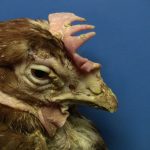
- In the chronic form, lesions are localized to wattles (facial edema), joints, sternal bursa, plantar pads, middle ear (may develop into torticollis), ovary, liver (perihepatitis), skull bones (fibrinous deposit) or respiratory system (infraorbital sinusitis, pneumonia, aerosacculitis, pneumatic bone disease).

The diagnosis
Clinical diagnosis
It is difficult. It can be suspected when high and sudden mortality affects birds of several species in a farm, especially when palmipeds are affected first. Autopsy cannot provide confirmation, even when staining on liver associated with heart and intestinal damage is observed.
Differential diagnosis
It concerns many conditions. A distinction must be made between highly pathogenic avian influenza (!) pasteurellosis, Newcastle disease, avian salmonellosis, duck plague, infectious rhinotracheitis (metapneumovirus infection), turkey erysipeloid, and all respiratory diseases, including infections caused by Avibactrium gallinarum and Gallibacterium anatis biovar haemolytica, which are related bacteria.
Laboratory diagnosis
P. multocida is isolated from bone marrow, liver, heart blood, localized lesions, swabs from nasal cavities. An antibiotic susceptibility test is often necessary to define the antibiotic susceptibility profile. Serological tests (ELISA) have limited value. At most, they are indicated for monitoring – roughly – the vaccination response.
Disease prevention and control
Treatment
If quickly implemented, it is effective in acute forms, but disappointing in chronic forms and too late in highly acute forms.
Antibiotic treatment is based on an antibiotic susceptibility test, combined with vitamins (A, B and C). Quinolones (nalidixic acid, oxolinic acid, flumequine, enrofloxacin), cephalosporins (ceftiofur), spectinomycin, amoxicillin (20 mg/kg PV), tetracyclines (doxycycline, chlortetracylcin at 40mg/kg), phenols (chloramphenicol at 20mg/kg) will be used mainly. The treatment is applied for at least 5 days, and must be adjusted according to the results of the antibiotic susceptibility test.
Health prophylaxis
It is difficult to implement. It consists in eliminating potential sources of P. multocida (sick or convalescent birds, rats, other birds,…), preventing contamination of food and drinking water, avoiding mixtures of species and age.
Medical prophylaxis
Consists of chemoprevention and/or vaccination. Chemoprevention may be recommended in recurrently affected farms. Vaccination is based on the use of inactivated agent vaccines. Commercial vaccines with the most common valences, or autovaccines, can be used.


