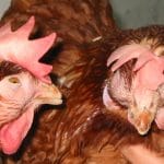
Avian mycoplasmoses are respiratory, genital or joint infections. These are insidious, common diseases, which have nevertheless declined in recent years, following eradication efforts in breeding flocks. They lead to heavy economic losses.
The disease agent and its pathogenicity
The etiological agent of mycoplasmosis is a mycoplasm. It is a small bacterium with a diameter of 0.2 to 0.5 µm, without walls. It is not visible in optical microscopy. Mycoplasmas are difficult to cultivate. They bind the red blood cells together.
Due to their lack of wall, mycoplasmas are resistant to many antibacterial agents, including b-lactam antibiotics. However, they are sensitive to most common disinfectants. Mycoplasmas can only survive for a few days outside their host.
There are many species, whose pathogenicity and host spectrum are variable. The main species of interest in avian pathology are : Mycoplasma gallisepticum (MG), M. meleagridis (MM) and M. synoviae (MS).
Epidemiological data
M. gallisepticum
Causes chronic respiratory disease in chicken and infectious sinusitis in turkey. MG has also been isolated from pigeons, ducks, geese, pheasants, partridges, quail and wild birds. Youngs are more sensitive than adults.
The infection is exacerbated by a previous bacterial or viral infection (Newcastle disease, infectious bronchitis, ILT), stress (vaccinations, other interventions…) and especially, by a bad environment (high NH3, dust).
Mycoplasmosis is often associated with other infections and/or favourable environmental conditions.
As the bacterium survives for a short time in an outdoor environment (1 to 3 days at 20°C), carrier birds are necessary for the sustainability of the disease. Transmission is vertical, and horizontal, direct (by infected youngs, wild birds) or indirect (environmental contamination). Backyards are often subclinically infected : they are a reservoir of the pathogen. In addition, multi-age breeding is a major risk factor.
M. meleagridis
Causes infection in turkeys. The germ has a tropism for the Fabricius bursa and causes immunosuppression. Youngs are more sensitive than adults.
The transmission can be vertical. It is also horizontal by the respiratory route : the infection then remains localized at the respiratory level most of the time. It can also be horizontal through the contaminated hands of the manipulator during sexing or artificial insemination. In carriers, the germ is isolated mainly in the genitals and sperm. Carriers often disappear after a breeding season.
M. synoviae
Causes infectious synovitis in chicken, turkey and guinea fowl.
Youngs are more sensitive than adults. Broilers are affected acutely, especially between 4 and 16 weeks of age, turkeys between 10 and 24 weeks of age. Chronic infection can develop and persist throughout the life of the batch. Cases are more frequent during cold and humid periods.
The bacterium is found in tendons, joints, ovaries and to a lesser degree in the respiratory tract.
The frequency of cases is currently decreasing. However, one clinical form is increasing : egg apical abnormality (also called glass top egg syndrome), which is characterized by fragile shells, especially at the apex of the egg, in laying hens.
Transmission is mainly vertical (transovarian), but also horizontal, via aerosols.
Clinical manifestations of the disease


M. gallisepticum
The incubation phase is from 6 to 21 days. Clinical signs often persist for a long time and are caused by a change. They are more severe in youngs and in turkeys.
Symptoms
- In turkeys, coughing, sneezing, rales, nasal and eye discharge, and swelling of the infra-orbital sinuses may occur (often, swelling is not associated with signs of deep respiratory system involvement).
- Chickens may have coughs, rails and nasal discharge, but this species is less sensitive to MG than turkeys. Clinical signs are more severe in youngs, but do not appear until 4 weeks of age.
- In laying hens, there is a decrease in food consumption and egg laying.
Lesions
Cachexia, catarrhal inflammation of the sinuses, trachea, bronchial tubes, opacification of air sacs with foamy or caseous exudate (chronic form), fibrinous pericarditis and perihepatitis, salpingitis (turkey).
M. meleagridis
NB : has become very rare in France.
Symptoms
Clinical signs are generally very weak. Egg hatchability decreases. Youngs have a lower growth rate. Sometimes turkeys have sinusitis or aerosacculitis, and the lesions often regress on their own. There are also deformations of the legs, and deforming osteomyelitis of the cervical vertebrae (turkeys with twisted necks).
Some turkeys show the following signs : poor growth and feathering, chondrodystrophy, aerosacculitis and diarrhea ; this is known as the “turkey syndrom 65”.
Lesions
Small amount of yellowish exudate in the air sacs (regressive lesions, often disappeared at the slaughterhouse) ; in “twisted neck” syndrome, turkeys show spondylitis and aerosacculitis in the cervical sac; in turkey syndrom 65, turkeys show chondrodystrophy, and a uni- or bilateral varus.
M. synoviae
Symptoms
Lameness, birds on the ground, pale ridges, swollen legs, growth delays, green droppings, generally asymptomatic respiratory infections.
Lesions
There is a viscous, grey to yellowish exudate in the joints (especially in the hock, wings, feet). During chronic infection, birds are emaciated, with an orange to brown dry exudate in the joints, as well as sternal bursitis (related to the friction of the furcula against the ground). Some birds, without joint damage, may have a slight tracheitis, sinusitis, aerosacculitis.
The diagnosis
Laboratory diagnosis
Serology is possible for MG and MS : agglutination tests are performed in tubes or on slides, and MG-MS is distinguished by inhibition of hemagglutination.
Culture is possible from orbital, nasal or tracheal swabs, MG tissues, embryos, tracheal, cloacal, vaginal swabs, phallus swabs for MM, joint swabs, spleen or liver samples in acute MS cases, lungs and air sacs in chronic cases. However, the growth of bacteria is slow, and can take up to 3 weeks. Diagnosis of mycoplasmosis by PCR is available on a routine basis, in particular with the help of commercial PCR kits.
M. gallisepticum
Clinical diagnosis
History of chronicity, weight loss, drop in egg laying, lesions.
Differential diagnosis
Colibacillosis, ORT, aspergillosis, avian cholera ; in turkeys, sinusitis can be caused by low pathogenic influenza viruses, M. synoviae.
M. synoviae
Clinical diagnosis
Lameness, swollen legs, lesions with grey to yellow exudate.
Differential diagnosis
Staphylococcal arthritis, viral arthritis, typhosis, pullorosis.
Disease prevention and control
Antibiotics are used to treat mycoplasmosis. Due to the absence of walls of these mycoplasma, antibiotics inhibiting wall synthesis (penicillin) and those inhibiting membrane synthesis are obviously ineffective. Several antibiotics inhibiting protein synthesis in combination (macrolides, doxycycline, 3rd generation quinolones) should be used. Antibiotics should be adapted to the resistance of the mycoplasmas involved. Antibiotic therapy should also help to combat frequent bacterial co-infections.
Eradication & prevention
Eradication & prevention
- Improve environmental conditions, pay particular attention to stress factors, ammonia levels and dust.
- Avoid the introduction of contaminated birds into an unscathed farm. The introduction of new animals must be from Mycoplasma spp-free breeding flocks ; the breeding animals are serologically monitored, their eggs are disinfected and can be treated, the chicks are raised in a sanitized and monitored environment. (Exported poultry must be certified free of MG and MM : this control applies particularly to trade in 1-day-old chicks).
Vaccination
- Vaccination against MG is also used in some countries, particularly in the Maghreb. Vaccines with inactivated agent are not very effective. Vaccines with attenuated live agent present a risk of reversion to virulence and make it difficult to identify contamination by a pathogenic wild isolate.
- Vaccination against MS, with a live attenuated agent vaccine, provides effective control of glass top egg syndrome.
Other mycoplasmas
M. iowae
It is mainly found in turkeys, but also in chickens and wild birds. The lesions are similar to those of M. meleagridis. It has become rare.
Duck mycoplasmas
These are mainly M. anatis, whose pathogenic role is still unclear. It could contribute to non-specific respiratory diseases of growing ducks.
Goose mycoplasmas
It is mainly M. anseris, its pathogenic role is obscure. A form of venereal mycoplasmosis in M. Anatis is specific to breeding animals : it causes very severe necrotic lesions of the penis in males and moderate cloacitis in females. Females are therefore mainly carriers, which contaminate gander. Penile necrosis in a significant number of gander is the call sign of this infection.


