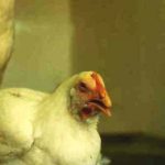
Coccidiosis is one of the most common parasitic diseases in poultry. They can take many forms and are found worldwide and in all types of poultry farming.
The disease agent and its pathogenicity
The etiological agent is a obligate protozoan intracellular parasite, most often belonging to the genus Eimeria. There are several species of coccidia for each bird species. The main species of coccidia of interest are the following :
- Chicken Coccidiosis :
A.acervulina, E. necatrix, E. maxima, E. brunetti, E. tenella, E. mitis, E. praecox - Turkey Coccidiosis :
E. meleagrimitis, E. adenoeides, E. dispersa, E. gallopavonis - Goose Coccidiosis :
E. truncata (pouvant aussi toucher le canard de Barbarie et le cygne), E. anseris - Duck Coccidiosis :
Tyzzeria perniciosa, E. mulardi (concernant principalement le canard mulard) - Guinea fowl Coccidiosis :
E. numidia, E. grenieri (plus fréquente mais au pouvoir pathogène inférieur) - Pigeon Coccidiosis :
E. labbeana
The cycle of coccidia is the same, regardless of the species of coccidia.
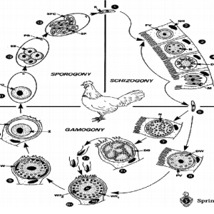
There are 2 phases of the life cycle : sexual and asexual. Asexual multiplication or schizogony takes place in the intestinal epithelial cells. Sexual multiplication or gamogonia results in fertilized eggs or oocysts, which are released into the intestine and then into the external environment. It is a direct monoxene two-phase cycle. The prepatent period (time between ingestion of the parasite and excretion of oocysts in the droppings) is 4 to 7 days.
Oocysts are highly resistant to most disinfectants and environmental conditions. They are the form of resistance of coccidia in the external environment
During the infestation of a batch of poultry, birds gradually become immune to coccidia, but there is no cross-protection against the different species of coccidia. Anticoccidians do not prevent the establishment of immunity because they do not destroy all coccidia but limit their load in the digestive tract. The acquisition of strong immunity is not an objective in broiler farming, due to their short lifespan.
Often, the host tolerates the moderately loaded parasite well, but all immunosuppression factors are favourable for disease expression. The damage caused is mechanical and is mainly located in the intestine.
Epidemiological data
There is a host specificity for each species of coccidia.
Young birds are more susceptible, especially broilers 3 to 6 weeks old and pullets. The disease is rare in layers and breeders. In turkeys, there are few signs after 8 weeks. However, the disease can appear at any age as a complication of another disease.
Coccidiosis is transmitted horizontally, directly from one bird to another of the same species through the faeces. It can also be transmitted indirectly by mechanical vectors (farm equipment) or insects (tenebrios).
Coccidia are ubiquitous in the environment.
Clinical manifestations of the disease
Clinical signs vary according to the species, infestation dose and degree of immunity of the bird: they can range from an inapparent form to loss of skin colour, growth retardation or reduced performance, prostration, and then diarrhea with dehydration and death.
-
Chicken coccidiosis
- E. acervulina : moderately pathogenic. The lesions are located in the small intestine mainly at the duodenum, with whitish spots and striae in the mucous membrane = “in scale” lesions. The lesions are caused by oocysts.
- E. necatrix : rare but highly pathogenic. The lesions are located at the end of the duodenum to the middle of the ileum. We have petechiae on the serosa (pepper and salt appearance) and whitish patches, mucus tinged with blood, distension of the intestine. The lesions are caused by 2nd generation schizonts. There is often a recrudescence between 9 and 14 weeks, because it is disadvantaged by competition with other coccidia. It is also called “chronic coccidiosis”.
- E. maxima : moderately pathogenic. The lesions are located from the end of the duodenum to the middle of the ileum. There is orange mucus and distension of the loops, thickening of the wall, petechiae, sometimes blood.
- E. brunetti : moderately to highly pathogenic. The lesions are located at the end of the small intestine and in the rectum. In severe cases, lesions may occur throughout the intestine, petechiae and mucosal necrosis, sometimes with blood and necrotic cylinders. The lesions are caused by schizonts.
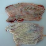
- E. tenella (cf. photographs 2 and 3) : the most pathogenic. The lesions are caused by schizonts and are located in the caeca, filled with blood, which can rupture or be gangrenous. The carcass may be anemic. Mortality is often high.
- E. mitis : low pathogenic. The lesions are in the second half of the small intestine. There are no macroscopic lesions, but mucus is present.
- E. praecox : low pathogenic. Mucus cylinders are noted in the duodenum. The prepatent period is short (83 hours).


- Turkey Coccidiosis :
Coccidiosis are common in turkey but are rarely diagnosed : the lesions are less dramatic than in chicken, and turkeys often heal quickly.
-
- E. meleagrimitis : the most pathogenic. The lesions are located in the first half of the small intestine. There is mucus and fluid in the duodenum with sometimes blood in the faeces.
- E. adenoeides : one of the most pathogenic. The lesions are mainly found in the caeca. Faeces are liquid, tinted with blood, sometimes with mucus cylinders.
- E. dispersa : low pathogenic. It is found in the small intestine. It is also isolated in quail, partridge and pheasant.
- E. gallopavonis : necrotic cylinders are found in the ileum and rectum.
- Goose Coccidiosis :
- E. truncata : mainly present in the kidney of goslings, it can cause high mortality on geese from 3 to 12 weeks old. The kidneys are enlarged, grey in colour, with small white foci and petechiae.
- E. anseris : present in the small intestine : mucus tinged with blood is observed. It can cause sudden death, with massive bleeding in the upper small intestine.
- Duck Coccidiosis :
They often cause bleeding damage : they can therefore cause sudden death, associated with massive bleeding in the small intestine.
-
- T. perniciosa : mainly located in the duodenal area, it can cause high mortality in youngs ; duodenal lesions are haemorrhagic with whitish spots visible on the serous side.
- E. mulardi : located in the jejuno-ileum and caeca, it can cause significant mortality with congestivohemorrhagic damage, especially in the jejuno-ileum, but may affect the entire intestine (except the duodenum).


- Guinea fowl Coccidiosis :
Primary coccidiosis is rarely observed in guinea fowl, which are quite tolerant of infestation. Moreover, the symptoms are not characteristic. Thus, most of the time, it is preferable to talk about the presence of oocysts rather than coccidiosis. Experimental inoculations have made it possible to reproduce signs in guinea fowl similar to coccidiosis in chicken, i. e. mortality, reduced performance. In addition, severe forms with bloody diarrhea have sometimes been encountered.
-
- E. numidia : is located in the small intestine and rectum. E. numidia infestations can cause mortality, with haemorrhagic diarrhea. They can result in a more or less important enteritis (slightly inflamed intestine, but without specific lesion).
- E. grenieri : is located in the intestine (schyzogonia) and the caeca and rectum (gametogonia). The disease mainly affects guinea fowl at 3 to 6 weeks of age, especially in winter ; the animals have diarrhea and the lesions are discreet. The infestation can cause enteritis in the duodenum and jejunum (corresponds to schizogony), or a slight liquefaction of the caecal contents (gametogony progression).
The diagnosis
Clinical diagnosis
It is difficult, due to unspecific symptoms and frequent co-infections. Lesions, if well marked, can be characteristic. Traditionally, coccidiosis lesions are graded at necropsy from +1 (mild) to +4 (severe).
The diagnosis is made by scraping the intestinal mucosa in various places and observing coccidia under a microscope between a slide and a cover slip. Eggs of E. brunetti, praecox, tenella and necatrix cannot be identified on the basis of oocyst size alone. Counting oocysts in the faeces makes it possible to monitor the evolution of contamination in a farm, but does not make it possible to manage the coccidial risk alone. A distinction must always be made between the carrying of coccidia and the clinical expression of coccidiosis.
Differential diagnosis
Necrotic enteritis, Non-specific enteritis, Histomoniasis.
Disease prevention and control
Medical prevention
Prevention involves the use of anticoccidial agents as additives or vaccination.
Several programs exist and must be defined, taking care to avoid the emergence of resistance :
- In broilers : use of the same molecule throughout the batch (continuous), or 2 molecules used successively in the same band (shuttle or dual program), or change of anticoccidian after a certain number of batches (rotation program).
- In layers and breeders : immunity is promoted by using live commercial vaccines, or anticoccidials are used, the dose of which is gradually reduced before laying.
- In turkey: anticoccidial agents are more toxic than in chicken, so they should be used with caution.
Prevention also involves the use of vaccination : live vaccines are registered in France and are based on early strains of major coccidial species (5 or 8 strains, depending on the Paracox 5® or Paracox 8® specialty). Vaccination gives good results and the use of these vaccines is now widespread on productions with high economic value (chicken labels, future breeders, etc…) which justify this cost of prophylaxis.
NB: the so-called “early” strains have the particularity of differentiating rapidly into male or female gamontes, after a small number of cycles of asexual division (schizogony) : parasitic cycles can therefore take place and generate a local immune response, without causing significant damage to the digestive mucosa.
Health prevention
Biosecurity in livestock farming is the only way to limit the risk of infestation or at least keep it below an equilibrium threshold :
- The control of oocyst entries from outside the shed limits contamination of the birds’ environment : boots or overboots, shed’s specific clothing, foot bath, clean and concrete access, control of wild animals, limitation of visits.
- A good cleaning and disinfection protocol at the end of the batch makes it possible to eliminate coccidia at the end of the breeding process and to start a new batch with a low parasitic pressure. Disinfection alone has no effect on oocysts.
- Limiting contact between birds and oocysts in faeces breaks the parasite cycle : use of cages, gratings, thick litter
- Bird health monitoring is important : coccidia are opportunistic parasites that take advantage of bird weakness to infest the host.
Treatment
Prevention measures do not always prevent the onset of the disease. Treatment should then be considered. The specialities used then comply with the legislation on veterinary medicinal products.
The treatment uses anticoccidials, synthetic products or ionophores : toltrazuril (BaycoxÒ), sulphonamides, amprolium (NémaprolÒ) in water or food.
In palmipeds, anticoccidial medication classically uses sulphonamides, amprolium (NemaprolÒ) and, above all, toltrazuril (BaycoxÒ). This prescription is done under the responsibility of the veterinarian…
- Parasitology website designed and maintained by Mr. JM Repérant (ANSES Ploufragan) : http://eimeria.chez-alice.fr/Accueil.html
- Diseases of Poultry, 12th Edition (2008). ed. Blackwell


